a-) Lasik Surgery

Lasik surgery is currently the most used procedure to correct definitively refractive defects such as myopia, hyperopia and/or astigmatism.
The Lasik operation is performed with a special instrument which is a microkeratome. The microkeratome cuts the cornea automatically the specialist can shape the tissue using the excimer laser. During Lasik surgery, any irregularities in the cornea or its curvature are fixed to ensure that the light entering the eye is projected correctly onto the retina. Usually, surgery takes about 15 minutes, and the procedure is completely painless and safe.
The Lasik technique has advantages in comparision to other refractive surgery procedures like;
The most widely used process in the world to correct vision defects.
Precise, successful, and safe surgery.
rapid recovery of visual acuity.
The postoperative is comfortable and painless the results can be noticed in 24 hours.
b-) Cataract Surgery ( Femtolaser/Femtosecond)

The most effective treatment in the cure of cataracts is performed by the surgical extraction of the opaque lens with aging and its replacement with an intraocular lens of synthetic material. (The lens is a natural lens located in the eye behind the iris, whose function is to focus light rays on the retina).. The highest technology of cataract surgery is the femtosecond laser or femtolaser.
The femtosecond laser machine has an infrared light with high-speed pulses the size focusing different layers in the eye to cut the desired area. Due to high speed, the laser light beam produces low energy to not damage the inside eye. It can be defined as robotic cataract surgery since it is the computer-guided laser that carries out the most delicate phases of the operation in an automated way.
The surgeon visualizes the procedure in real-time on a monitor which projects an image of the section of the eye to be treated. The femtolaser can be used to improve the safety and precision of cataract surgery.. The laser beam is considerable: the cuts made on the cornea to then remove the crystalline lens are very precise and small in size and allow for the reduction in the onset of astigmatism postoperative corneal, also guaranteeing rapid visual recovery times and quality of vision. The correction of an already existing astigmatism can also be performed by the laser machine, creating corneal cuts of the desired length and depth, depending on the patient`s astigmatism.
The use of the femtolaser in cataract surgery aims to make some surgical steps safer, more precise, repeatable, and reproducible by carrying out the most delicate phases of the operation in an automated way.
c-) Vision correction (PRK laser)

PRK or photorefractive keratectomy is an eye treatment that is used to treat defects of myopia, hyperopia, astigmatism, and presbyopia. The PRK technique was performed first time in the early 1980s, then in 1995 that it was approved by the FDA.
With PRK the cornea of the eye was exposed. Thus it is to remove the corneal epithelium by the diluted alcohol,.This alcohol weakens the epithelium so that later, with a spatula, it can be completely separated and thus subsequently apply the excimer laser to the stroma. With the help of an excimer laser, a new surface of the cornea is formed. The course of laser correction is controlled by an ophthalmic surgeon. After the laser correction is completed, the cornea is washed with a special solution. The patient is given anti-inflammatory drops. Surgery is usually performed under local anesthesia. The surgery is performed on both eyes instantly surgical act and the approximate time of the entire process is about ten minutes.
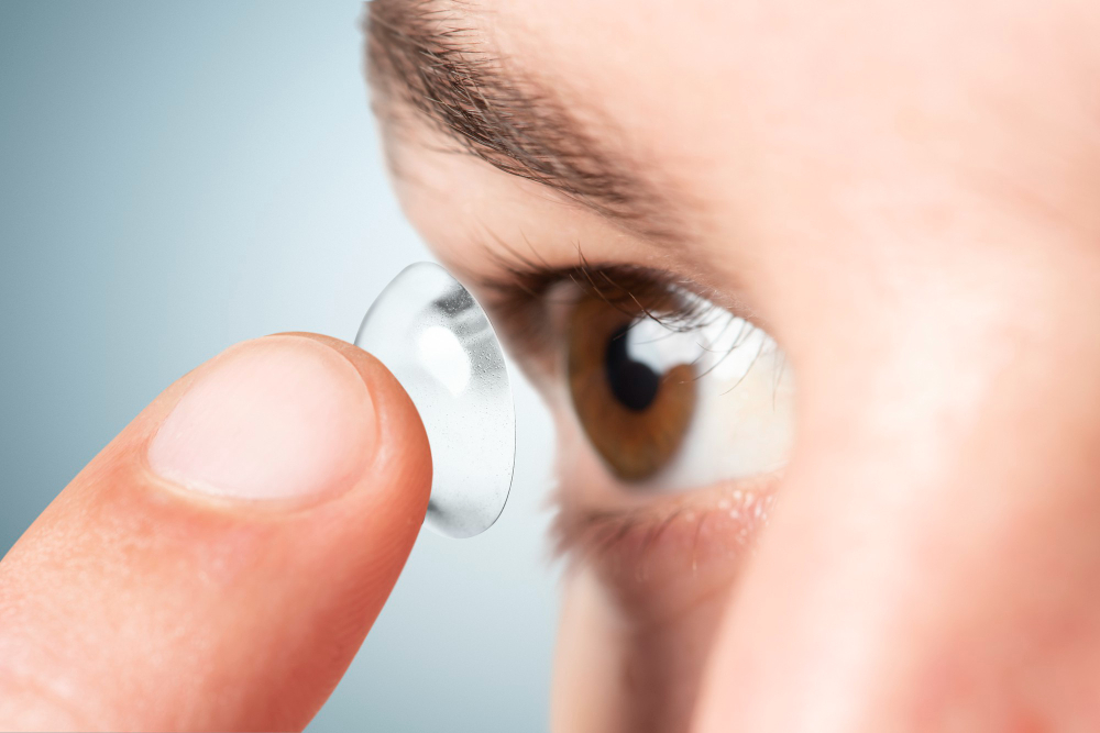
After the surgery, it is placed soft "band-aids" like contact lenses on the eyes for protection during healing time between 3-7 days until the epithelium is regenerated again. Just after the surgery, the patients turn back to their daily life and reading activities, and full adoption time ends in 3-4 weeks.
Candidates for PRK
Patients with dry eye who have been using contact lens users for many years and who no longer tolerate them.
Patients with thin corneas. With Lasik treatment, the laser s is evaporated stromal tissue, and the greater the amount of diopters, the more amount of tissue it is evaporated, therefore, if the cornea is too thin, it is not preserved the adequate margins by Lasik treatment.
Patients who practice contact sports with hit risk in the eye or have jobs Since, if a LASIK is performed when receiving the blow, the pocket we made could be displaced. On the other hand, in the PRK technique, since there is no pocket, it cannot be moved.
Patients who have myopia from -1.0 to -6.0 D and astigmatism from -0.5 to -3.0 D.hyperopia up to +3.0 D are not good canditates for PRK .
d-) Corneal Transplantation(Keratoplasty)

Corneal transplantation or keratoplasty is a surgical procedure that consists of replacing damaged corneal tissue with healthy corneal tissue from a donor when there is irreversible corneal alteration that can not be corrected in any other way. This transplant surgery is the most frequently performed worldwide. It is a transfer surgery that is applied to patients who have transparency is impaired or the ultra-showing deviation of curvature of the cornea tissue.
Cornea transplantation is performed only with tissue taken from the dead body.So,a healthy person can not donate his cornea even if he is twin of the patient.The cornea is one of some tissues that can be alive for a while after death. Just after the deah, the cornea is taken and transferred to the transplant center in special storage solutions, and its vitality is preserved for a while.
After examination by ophthalomogist the patients who are examined by the Ophthalmologist and who are considered to have corneal transplantation are admitted by their physicians to Turkish Eye Bank with patient identification and clinical information and a patient record is created from the registry of the Ministry of Health.
e-)Squint ( Strabismus)

Strabismus is the loss of alignment of the visual axes, either in all visual directions or in one of them means that both eyes cannot focus on the same point simultaneously.
Strabismus İS an aesthetic or functional problem. The problem can be a residual condition from childhood, or due to a process acquired in adulthood (associated with diseases such as strokes, tumors, thyroid ophthalmopathy, or aging), in which case they can go accompanied by diplopia (double vision), a condition that can be very limiting in daily life for activities such as driving, reading or watching television, as well as disabling for many jobs.Usually, the disease appears in early childhood – at 2-3 years
Strabismus should be evaluated by a specialized ophthalmologist, who will evaluate the different treatment possibilities to improve both aesthetics and visual function as far as possible. Sometimes a similar disease develops – amblyopia ("lazy eye"), when the squinting eye is partially or completely excluded from the visual process.
There are different types of strabismus:
Converging strabismus (esotropia) – the eye deviates to the bridge of the nose; the condition is often accompanied by farsightedness.
• Divergent strabismus (exotropia) – the eye is tilted to the temple; the condition can be triggered by myopia.
• Vertical strabismus – the eye is tilted up (hypertropia) or down (hypotropia).
• Mixed strabismus – combines several forms at once.
The first step consists of a correct diagnosis of the type of strabismus and its consequences (amblyopia, diplopia, etc.), to then focus on the treatment of both.
Each case must be individualized, taking into account the type of strabismus, the age at diagnosis, the depth of the lazy eye, the necessary graduation, and other characteristics that may lead us to opt for a different combination of treatments.
Likewise, the collaboration of the families is important, since the process is usually long and requires a significant effort on the part of the parents in terms of time and patience. Surgical treatment corrects this defect.
During strabismus surgery, the muscles that move the eyeball can become weaker or stronger. The position of the muscles can also be moved. To weaken the muscle, a suture is placed in the muscle. The eyeball muscle is then removed and reattached further back. To strengthen the muscle, the muscle is shortened to increase its ability to pull the eye.
The eyes always move together. Although it appears that either the right or left eye is misaligned, the problem is the balance between the eyes. Therefore, surgery can be performed on one eye or both eyes to balance and straighten the eyes. Strabismus surgery is performed on an outpatient basis, usually under general anesthesia. During surgery, incisions are made in the conjunctiva. The conjunctiva is the thin covering over the eye. In children, the incisions are usually made in the pocket between the eyelid and the eyeball to hide the scars.
In older patients and patients with previous surgeries, incisions can be made where the iris touches the white part (the limbus). In most cases, the scar is not visible. After surgery, the white of the eye will be very red in the incision area. This is a bloodstain. The bloodstain will dissolve in about two weeks. However, the white part of the eye may remain inflamed and red for several more weeks. In some cases, particularly if there are scars from previous surgery, the area may always remain a bit red. If the surgery is performed on both eyes, patches will not be placed on both eyes.